4DMedical Expands into Europe with Agreement to Acquire Software Lung Imaging Leader contextflow
4DMedical, a global leader in advanced respiratory imaging and lung analysis software, has signed a binding agreement to acquire contextflow GmbH, a Vienna-based medical software imaging company specializing in lung cancer detection, chest CT analysis and radiology workflow solutions. The acquisition establishes 4DMedical’s on-the-ground European commercial platform, adding an experienced regional team, an existing customer base, CE-marked products and MDR-aligned regulatory capabilities.
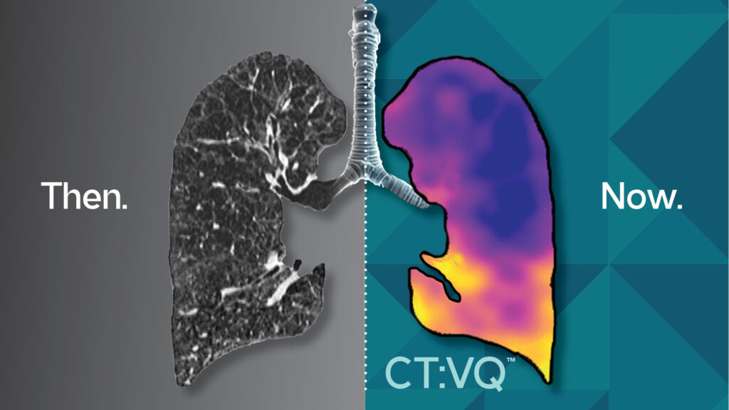
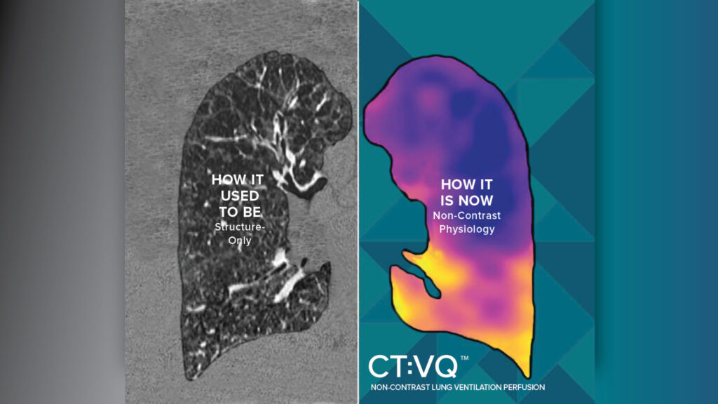
By combining contextflow’s software-driven structural lung imaging tools with 4DMedical’s functional lung imaging technologies, including XV Technology®, CT LVAS™ and CT:VQ™, the combined portfolio will support a more complete clinical pathway for lung disease detection, diagnosis, monitoring and disease progression tracking. The transaction strengthens 4DMedical’s global footprint across North America, Europe and Australia, accelerating commercialization, clinical validation and adoption of advanced lung imaging solutions worldwide.
4DMedical receives CE Mark for CT:VQ™, enabling commercial launch across the European Union
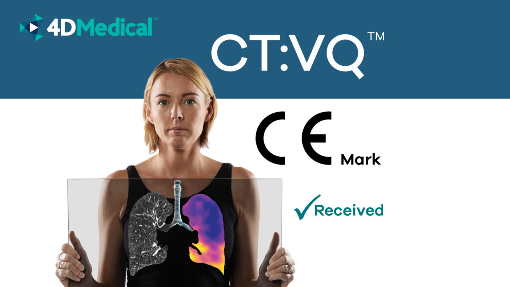
4DMedical Limited (ASX:4DX), the global leader in cardiothoracic imaging software, announces that its latest imaging technology, CT:VQ™ has received CE Mark certification for commercial use in the European
Union. 4DMedical will quickly launch commercial deployment of CT:VQ across one of the world’s largest respiratory imaging markets.
Not Just for Smokers: The Surprising Faces of Lung Disease

We may all be guilty of picturing the same person when we hear that someone has lung cancer—an older man who smoked for many years. The truth is, millions of people with lung disease have never smoked a single cigarette. Children, women who have never smoked, and even athletes can suffer from life-altering respiratory disorders, too.
Lung Imaging in 2026: How Functional CT and New Investments Are Changing the Game
Lung disease is a global health challenge. Conditions like COPD, emphysema, and lung cancer affect hundreds of millions of people worldwide, yet many patients are still being diagnosed too late. Traditional imaging has focused on structure: identifying nodules, masses, or obvious tissue changes. In 2026, the field is rapidly evolving toward functional insights, understanding not only what the lungs look like but also how they function, region by region. Recent research, industry debate, and major investment activity all point to a pivotal moment in pulmonary imaging.
Aunt Minnie: 4DMedical inks major distribution agreement for CT:VQ™

New coverage from AuntMinnie.com: Philips will distribute 4DMedical’s CT:VQ™ across healthcare systems in the U.S. and Canada, expanding access to non-contrast CT-based ventilation and perfusion mapping.
Catching COPD Early: Why Timing Could Save Your Lungs
Chronic Obstructive Pulmonary Disease, or COPD, is one of the most common lung conditions in the world—yet it remains deeply underdiagnosed. In the United States alone, over 12.5 million people are known to have COPD, but millions more may be living with it. One of the main reasons for this gap is that early symptoms, such as a lingering cough or shortness of breath, are often easily dismissed. Many people don’t seek help until their breathing problems are severe and irreversible damage has already taken place.
CT:VQ™ Redefines Lung Imaging: FDA Clearance Makes Non-Contrast V/Q Scans a Reality
In a landmark move for pulmonary diagnostics, 4DMedical’s CT:VQ™ earned FDA 510(k) clearance in September 2025, marking it as the first and only non-contrast ventilation–perfusion (V/Q) imaging solution. The technology is available via routine chest CT scans. With CMS confirming reimbursement under Category III CPT codes (on top of the existing chest CT payment), CT:VQ™ is poised to redefine accessibility and efficiency in functional lung imaging.
4DMedical’s CT:VQ™ Receives FDA 510(k) Clearance; First-and-Only Non-Contrast VQ Imaging
4DMedical, a leader in advanced respiratory imaging, today announces U.S. Food and Drug Administration (FDA) 510(k) clearance for CT:VQ™, the world’s first and only non-contrast, ventilation–perfusion (VQ) imaging solution. In parallel, the U.S. Centers for Medicare & Medicaid Services (CMS) has confirmed reimbursement for CT:VQ under Category III CPT codes; this payment is in addition to existing reimbursement for the underlying chest CT.
Understanding Lung Perfusion: What is it? Why does it matter? And how is it measured?

Lung health is essential for overall wellbeing. A key aspect of healthy lungs is their ability to efficiently transfer oxygen into the blood. This process relies on both the flow of air (ventilation) and the flow of blood (perfusion) through the lungs. While ventilation is about how well air moves in and out, perfusion refers to the movement of blood through the lungs’ fine network of blood vessels. Both are crucial, but today we’ll take a closer look at lung perfusion: what it is, when and why it’s measured, and how advanced imaging technologies are enhancing our understanding of it.
Addressing the Challenge of UIP—and IPF Diagnosis: A Conversation with Dr. Chung

Early identification of Usual Interstitial Pneumonia (UIP) and diagnosis of Idiopathic Pulmonary Fibrosis (IPF) remains a significant challenge in pulmonary medicine. Despite advances in imaging and clinical awareness, the process still requires specialized training and experience that isn’t always readily available—especially in non-urban or resource-limited healthcare settings. This gap can delay diagnosis which critically impacts patient outcomes.
In a recent interview hosted by Brian Casey, editor at The Imaging Wire, Dr. Chung, a leading thoracic radiologist, offers valuable perspective on the complexities associated with UIP identification and the essential role of expert interpretation to achieve accurate diagnoses.