Over 12.5 million Americans live with COPD. The discomfort caused daily by COPD increases rates of anxiety and depression in individuals and can severely limit their ability to work, causing significant financial damage to families. The American Lung Association estimates that the total economic cost of COPD is close to $50 billion each year, including $29.5 billion for direct healthcare expenditures, $12.4 billion for indirect mortality costs, and $8.0 billion for indirect morbidity costs.
Chronic Obstructive Pulmonary Disease (COPD) is a progressive lung disease that affects millions of people worldwide. It is characterized by airflow limitation and inflammation in the lungs, leading to symptoms such as shortness of breath, coughing, and wheezing. While COPD is a chronic condition that cannot be cured, early detection and management can significantly improve a patient’s quality of life. One crucial aspect of COPD management is utilizing lung imaging techniques to assess the extent of lung damage and monitor disease progression.
What is COPD?
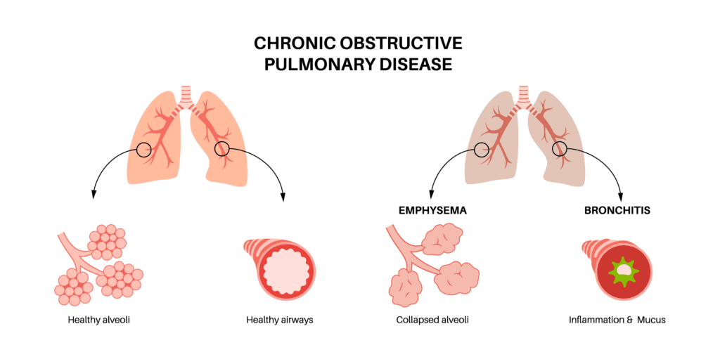
COPD stands for chronic obstructive pulmonary disease and includes a group of diseases including chronic bronchitis and emphysema that make it difficult for individuals to breathe comfortably. COPD is a chronic disorder meaning it is lifelong and while not entirely treatable, symptoms can be managed. The American Lung Association describes how COPD affects both the flow of air in and out of the lungs as the airways become swollen. The walls between the air sacs may be destroyed, and the airways and air sacs are no longer flexible and able to stretch and shrink as they would in healthy lungs. As COPD symptoms worsen, patients might also cough frequently with mucus, and as less oxygen enters the lungs, less carbon dioxide is exchanged, causing increased difficulty with breathing.
What are The Symptoms?
COPD symptoms typically start around age 40 or older. Individuals may excuse their symptoms as just a part of “getting older,” however, shortness of breath and a regular cough should always be brought to the attention of your physician. Common symptoms include:
- Shortness of breath
- Frequent cough with mucus
- Chest tightness and wheezing
- Feeling tired from an inability to breathe properly
- Frequent lung infections like pneumonia
What are the Common Causes?
Data analysis by the American Lung Association’s Epidemiology and Statistics Unit shows that smoking is the greatest risk factor—with smokers having a seven times higher rate of COPD than those who do not smoke. Additionally, exposure to certain environmental toxins may damage the integrity of the lungs putting members of the military, law enforcement, firefighters, and other workers at a higher risk. It is common to see higher rates of COPD in more rural and industrial parts of the country due to higher smoking rates, increased pollution, and jobs in toxin-producing industries, like coal mining. For example, the rate of COPD in West Virginia is double that of Hawaii.
Individuals with asthma or a history of severe childhood respiratory infections may also be at risk. Additionally, any serious respiratory infection like those caused by the Flu or COVID-19 increases the chance of pneumonia which can cause permanent lung damage and lead to COPD as well, especially in older or already at-risk individuals. Quitting smoking as soon as possible, avoiding environmental triggers, and keeping up with vaccinations are all recommended preventive measures.
Why are Women Uniquely Affected by COPD?
Today, more women than men have COPD in the United States. COPD was previously thought of as a “man’s disease,” and women are still underdiagnosed which can lead to worsening symptoms and mismanagement of care. The tobacco industry heavily targeted women in the 1960s, many of whom are now experiencing COPD symptoms after a lifetime of smoking.
However, fewer women than men with COPD were smokers, and researchers speculate that the smaller size of female lungs and estrogen may also play a role in the disease.
How is COPD Diagnosed?
Traditionally, COPD has been diagnosed based on symptom reporting and spirometry, a simple test that measures lung capacity. Lung imaging plays a vital role in the management of COPD by facilitating early detection, accurate diagnosis, disease severity assessment, and ongoing monitoring. Healthcare providers should integrate imaging studies into COPD care protocols to optimize outcomes and improve patients’ quality of life. There are several imaging modalities used in the evaluation of COPD:
Chest X-rays: X-rays provide a two-dimensional image of the lungs and can help identify abnormalities such as lung hyperinflation, consolidation, or the presence of other lung conditions such as pneumonia. However, X-rays may not detect early signs of COPD and are less sensitive than other imaging techniques.
Computed Tomography (CT) scans: CT scans offer a more detailed view of the lungs when compared to X-rays. They can detect emphysema, bronchiectasis, and other structural changes associated with COPD. CT imaging is particularly useful for assessing the severity of emphysema and determining the presence of bronchial wall thickening or bronchiectasis.
Magnetic Resonance Imaging (MRI): While less commonly used and more costly than X-rays and CT scans in COPD diagnosis, MRI can provide valuable information about lung perfusion and tissue characteristics. It is useful for evaluating pulmonary vascular changes and detecting early signs of pulmonary hypertension, a complication of advanced COPD.
Emerging, non-invasive tests are the new frontier in diagnosis. Investigators from Johns Hopkins and the University of Miami presented findings from their COPD study at the International Conference of the American Thoracic Society and found that 4DMedical’s XV Lung Ventilation Analysis Software was capable of assessing regional ventilation defects, which is critical to optimizing therapies.
4DMedical’s XV LVAS Technology takes a unique approach to imaging lung function, using existing hospital imaging systems. Fluoroscopy and CT scans are analyzed using image-processing methods adapted from aerospace engineering. Algorithms measure the motion of the lung tissue. From motion, ventilation is calculated at each stage of the breath and every location within the lung. The ventilation measurements are then visualized as a colored heat map to identify ventilation deficits. The software provides radiologists and physicians with a report highlighting regional ventilation, lung volumes, heterogeneity, etc.
What Treatments Are Available?
COPD treatments fall into several categories, the first being non-invasive which includes
- Quitting smoking
- Pulmonary rehabilitation combines exercise training, disease management education, social support, nutrition education, and counseling.
- Medications like bronchodilators to relax the airways, steroids, antibiotics when needed for infections, and vaccinations to prevent serious illness.
- Supplemental oxygen therapy
- Non-invasive ventilation is a form of noninvasive positive pressure ventilation (NPPV). This may help prevent hospitalizations and reduce mortality in individuals with higher levels of carbon dioxide in the blood. It can be administered through a simple face mask at home.
Open surgical options come with many serious risks, and many patients are not candidates. They include,
- Lung transplants
- Lung volume reduction surgery to remove diseased tissue
- Bullectomy to remove damaged air sacs
New, non-surgical treatments.
EBV therapy is a new, minimally invasive procedure where a one-way valve is placed that reduces lung hyperinflation by allowing the trapped air to escape. Although it can’t cure COPD, it can greatly improve lung function and reduce shortness of breath without the risks of traditional open surgery.
As we continue to advance in our understanding of COPD and refine diagnostic and treatment modalities, collaboration among healthcare professionals, researchers, technology innovators, and policymakers becomes paramount. By leveraging the power of lung imaging alongside holistic care strategies, we can strive towards better outcomes, enhanced patient well-being, and ultimately, a brighter future in the fight against COPD and respiratory diseases.