Since 2006, our preclinical and clinical trials have consistently demonstrated that XV Technology provides an unprecedented level of information on respiratory function.
Our ground-breaking work has been published in more than 80 scientific publications.
Studies include information about X-ray velocimetry and lung heterogeneity. Lung heterogeneity may be a key indicator of lung health.
Quantifying ventilation by X-ray velocimetry in healthy adults
Association of X-ray velocimetry (XV) ventilation analysis compared to spirometry
Functional imaging for assessing regional lung ventilation in preclinical and clinical research
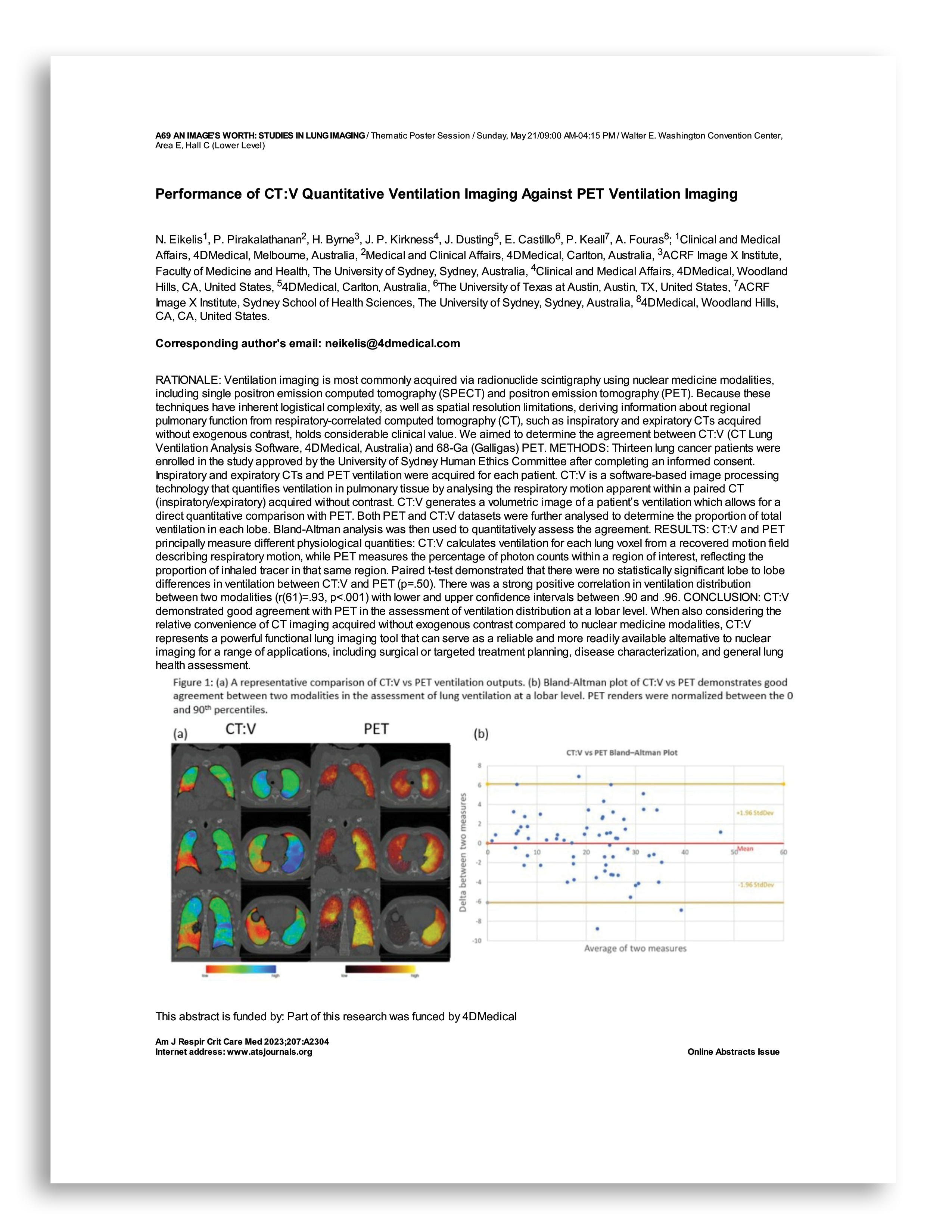
Performance of CT:V Quantitative Ventilation Imaging Against PET Ventilation Imaging
Hatt CR, Oh AS, Obuchowski NA, Charbonnier JP, Lynch DA, Humphries SM. Comparison of CT Lung Density Measurements between Standard Full-Dose and Reduced-Dose Protocols. Radiology: Cardiothoracic Imaging. 2021 Apr 1;3(2):e200503.
McEvoy CE, Lando-King E, Keith LA, Hatt CR, Arenberg DA, Kazerooni EA, et al. Development and impact of a patient-centered, CT image data-enhanced lung cancer screening CT report on smoking cessation behaviors. Chest. 2022 Oct;162(4):A2700–1.
Labaki WW, Xia M, Murray S, Hatt CR, Al-Abcha A, Ferrera MC, et al. Quantitative Emphysema on Low-Dose CT Imaging of the Chest and Risk of Lung Cancer and Airflow Obstruction.
Amundson WH, Swanson EJ, Petersen A, Bell BJ, Hatt C, Wendt CH. Quantification of Perinodular Emphysema in High-risk Patients Offers No Benefit in Lung Nodule Risk-Stratification of Malignancy Potential. Journal of Thoracic Imaging. 2020 Mar;35(2):108–14.
Gunning SG, Graby J, Mody Y, Charters PF, Burnett TA, Murphy D, et al. Visual ordinal grading of aortic valve calcification on routine non-gated chest CT predicts prognosis and alters management. European Radiology. 2025;1–11.
Foley RW, Glenn-Cox S, Rossdale J, Mynott G, Burnett TA, Brown WJH, et al. Automated calculation of the right ventricle to left ventricle ratio on CT for the risk stratification of patients with acute pulmonary embolism. Eur Radiol. 2021 Aug;31(8):6013–20.
Wang JM, Bose S, Murray S, Labaki WW, Kazerooni EA, Chung JH, et al. Quantitative CT Measures of Lung Fibrosis and Outcomes in the National Lung Screening Trial. Annals ATS. 2025 Apr 10;AnnalsATS.202410-1048OC.
Chung JH, Chelala L, Pugashetti JV, Wang JM, Adegunsoye A, Matyga AW, et al. A Deep Learning-Based Radiomic Classifier for Usual Interstitial Pneumonia. CHEST. 2024 Feb;165(2):371–80.
Wu JKY, Ma J, Nguyen L, Dehaas EL, Vasileva A, Chang E, et al. Correlation of respiratory oscillometry with CT image analysis in a prospective cohort of idiopathic pulmonary fibrosis. BMJ Open Resp Res. 2022 Apr;9(1):e001163.
Romei C, Falaschi Z, Danna PSC, Airoldi C, Tonerini M, Rocchi E, et al. Lung vessel volume evaluated with CALIPER software is an independent predictor of mortality in COVID-19 patients: a multicentric retrospective analysis. Eur Radiol. 2022 Jun;32(6):4314–23.
Romei C, Castellana R, Conti B, Bemi P, Taliani A, Pistelli F, et al. Quantitative texture-based analysis of pulmonary parenchymal features on chest CT: comparison with densitometric indices and short-term effect of changes in smoking habit. Eur Respir J. 2022 Oct;60(4):2102618.
Occhipinti M, Bosello S, Sisti LG, Cicchetti G, De Waure C, Pirronti T, et al. Quantitative and semi-quantitative computed tomography analysis of interstitial lung disease associated with systemic sclerosis: A longitudinal evaluation of pulmonary parenchyma and vessels. Kuwana M, editor. PLoS ONE. 2019 Mar 12;14(3):e0213444.
Nair A, Mohan R, Greeshma MV, Benny D, Patil V, Madhunapantula SV, et al. Artificial Intelligence Unveils the Unseen: Mapping Novel Lung Patterns in Bronchiectasis via Texture Analysis. Diagnostics. 2024 Dec 21;14(24):2883.
McCarthy C, Lee E, Bridges JP, Sallese A, Suzuki T, Woods JC, et al. Statin as a novel pharmacotherapy of pulmonary alveolar proteinosis. Nat Commun. 2018 Aug 7;9(1):3127.
Ferrazza AM, Gigante A, Gasperini ML, Ammendola RM, Paone G, Carbone I, et al. Assessment of interstitial lung disease in systemic sclerosis using the quantitative CT algorithm CALIPER. Clin Rheumatol. 2020 May;39(5):1537–42.
We use cookies to improve your experience on our site. By using our site, you consent to cookies.
Manage your cookie preferences below:
Essential cookies enable basic functions and are necessary for the proper function of the website.