SaaS Software

CT:VQ™

Maximum CONTRAST. No INJECTIONS.

Non‑contrast ventilation and perfusion from standard chest CTs
CT:VQ™ is the world’s first non‑contrast post‑processing technology that transforms routine, chest CTs into quantitative, lobar ventilation (V) and perfusion (Q) maps—without injected contrast or radioisotopes. Delivered via software-as-a-service, results are returned directly into the radiology workflow for interpretation alongside the source CT images.

FDA 510(k) Cleared, For Investigational Use Only outside of the United States.
Eligible for reimbursement under Category III CPT codes.

Designed for you

  • Pulmonologists and Interventional Pulmonologists: Obtain regional V/Q to support PE workups, CTEPH assessment, regional detection of obstructive/restrictive V/Q abnormalities, BLVR planning, pre‑op assessment, and longitudinal monitoring in a single-page report designed to support patient communication, alongside the radiology report.

  • Radiologists:  Add functional interpretation to non‑contrast chest CTs with structured, quantitative outputs delivered to PACS with DICOM Structured Reporting to minimize workflow while maximizing clinical benefits.

  • Hospital and Imaging Administrators: Expand access to V/Q imaging across existing CT infrastructure with no new hardware and streamlined scheduling. Reduce operating costs and gain reimbursement.

Breakthrough Features

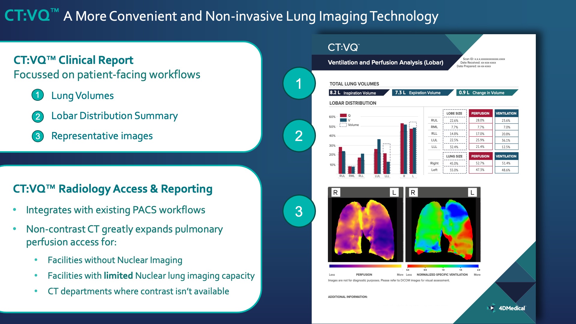
  • Color V and Q overlays registered to CT anatomy

  • Quantitative Metrics

  • Total Lung Volumes (inspiratory, expiratory, and delta)

  • Lobar Distribution: comparisons between lobe size, ventilation, perfusion, and total right–left lungs
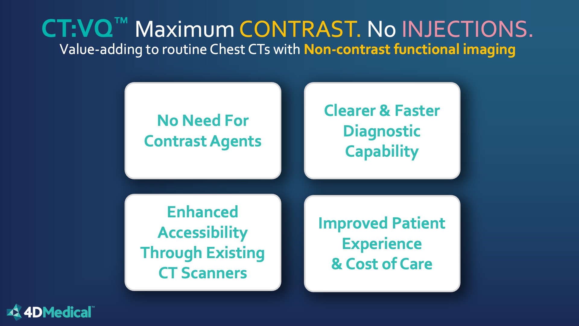
  • Structured reports with patient‑facing clinical workflows

Key Advantages

  • No Injections: Non‑Contrast CT only—no IV access, no radioisotopes, no contrast agents.

  • Clarity and Quantitation: High‑resolution, color‑coded functional maps with structured, lobar metrics.

  • Accessible at Scale: Runs on the approximately 14,500 CT scanners installed across the U.S.—bringing V/Q to sites without nuclear medicine capacity or where capacity is constrained.

  • Patient‑ and Operations‑Friendly: Single CT appointment; simplified scheduling; avoids contrast media, IV placement, and related consumables; supports review of prior CTs to help avoid repeat imaging. comparisons between lobe size, ventilation, perfusion, and total right–left lungs

The Non-Contrast CT:VQ™ Value-Added Solution

CT:VQ™—Non‑contrast Functional Lung Imaging, No Injections

Add functional insights to routine chest CT using existing scanners and PACS—eliminating contrast agents and supporting broader, patient‑friendly access.

Clinical Evaluation

CT:VQ™

CT:VQ™ was evaluated using three complementary approaches:
Data on file, 4DMedical

1. Quantitative correlation with SPECT V/Q (standalone device performance)

        • Lobar perfusion correlation: r = 0.872
        • Lobar ventilation correlation: r = 0.842 Notes: Each point represents a lobe’s percentage of total lung perfusion or ventilation.


        Additional Analysis: CT:VQ™ perfusion heterogeneity metrics showed a stronger association with DLCO than comparable SPECT measures in the submission dataset (r²: 0.557 vs 0.380).


        2. Reader performance study

        • Expert radiologists and nuclear medicine physicians graded regional function on CT:VQ™ and SPECT V/Q using a standardized 5‑point scale.
        • Agreement with SPECT V/Q was rated good to excellent across all lung zones.
        • Kendall’s tau by zone (all readers): 0.702–0.765, exceeding the pre-specified 0.40 target for the submission.
    •  

      3. Case‑based review

      • Illustrative cases demonstrated clinical interpretability across multiple conditions, highlighting areas of agreement and explaining discrepancies (e.g., SPECT ventilation limited by radiotracer deposition artifacts).

Get Started

  • Request a clinical demo and sample report

  • Submit a test case from your PACS

  • Plan a pilot across your CT fleet

What's new at 4DMedical

Get the latest news about respiratory imaging and ventilation analysis