EDUCATION HUB

Case Studies

Lorem ipsum dolor sit amet, consectetur adipiscing elit, sed do eiusmod tempor incididunt ut labore et dolore magna aliqua. Ut enim ad minim veniam, quis nostrud exercitation ullamco laboris nisi ut aliquip ex ea commodo consequat. Duis aute irure dolor in reprehenderit in voluptate velit esse cillum dolore eu fugiat nulla pariatur. Excepteur sint occaecat cupidatat non proident, sunt in culpa qui officia deserunt mollit anim id est laborum.

Case Study 1: Silicosis

AGE
45
Sex
Male

INDICATIONS

  • Biopsy-proven silicosis
  • Ex-Smoker
  • Subjectively, right-sided chest discomfort with breathing
SUMMARY
  • Unable to complete spirometry
  • Strong pain on the right side
  • LVAS demonstrated a functional difference in ventilation on the right side

Add Your Heading Text Here

Lorem ipsum dolor sit amet, consectetur adipiscing elit, sed do eiusmod tempor incididunt ut labore et dolore magna aliqua. Ut enim ad minim veniam, quis nostrud exercitation ullamco laboris nisi ut aliquip ex ea commodo consequat. Duis aute irure dolor in reprehenderit in voluptate velit esse cillum dolore eu fugiat nulla pariatur. Excepteur sint occaecat cupidatat non proident, sunt in culpa qui officia deserunt mollit anim id est laborum.

Case Study 2: Non-Obstructive Atelectasis

AGE
69
Sex
Female

INDICATIONS

  • Past history of Covid-19
  • Gastric Oesophageal Reflux Disease
  • Recurrent chest infections
SUMMARY
  • Imaging showed a large hiatus hernia
  • LVAS demonstrated a greater functional reduction in ventilation than expected from CT changes

Add Your Heading Text Here

Lorem ipsum dolor sit amet, consectetur adipiscing elit. Ut elit tellus, luctus nec ullamcorper mattis, pulvinar dapibus leo.

Case Study 3: COPD With Biologic Therapy

AGE
60
Sex
Female

INDICATIONS

  • Prior biologics therapeutic for re-current exacerbation of moderate obstructive lung disease.
  • Medication: Fasenra (benralizumab)
SUMMARY
  • SOB for further investigation.
  • At baseline CT was unremarkable. Placed on biologics for history of exacerbation.
  • Following Tx, there are functional improvements in regional ventilation indices (reduced VH and VDP). Notably, appearance of improved ventilation, specifically in the dependent areas of the right and left lungs.
  • Corresponding with patient reported improvement in symptoms.

Add Your Heading Text Here

Lorem ipsum dolor sit amet, consectetur adipiscing elit. Ut elit tellus, luctus nec ullamcorper mattis, pulvinar dapibus leo.

Case Study 4: Long COVID-19 Symptoms

AGE
52
Sex
Female

INDICATIONS

  • Long COVID-19 symptoms.
SUMMARY
  • Patient hospitalised for COVID-19.
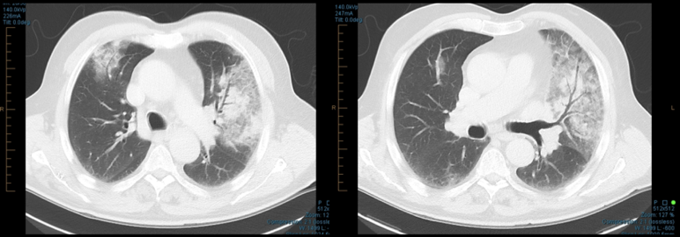
  • During hospital admission chest CT observed peripheral ground-glass and consolidative pulmonary opacities. (no XV LVAS imaging was captured)
  • Following Tx and discharge from hospital, the Pt continued to display symptoms of shortness of breath, cough and dyspnoea on exertion.
  • Following a physician consultation, the patient was referred for follow up CT and 4DMedical XV LVAS imaging.

Clinical Observations

XV LVAS:
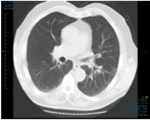
Follow up CT observed a resolution of the peripheral ground-glass and consolidative pulmonary opacities.
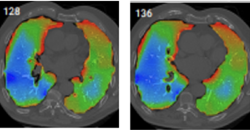
XV LVAS highlighted heterogeneity between the left and right lung regional performance.
Additionally, previous areas of ground-glass and consolidative pulmonary opacities displayed under ventilation specific to that region of the lung.
Chest CT: Acute COVID-19 Infection
Structural CT: Peripheral ground-glass and consolidative pulmonary opacities
3 Months Post-acute COVID-19 Infection
Structural CT
XV LVAS

What's new at 4DMedical

Get the latest news about respiratory imaging and ventilation analysis