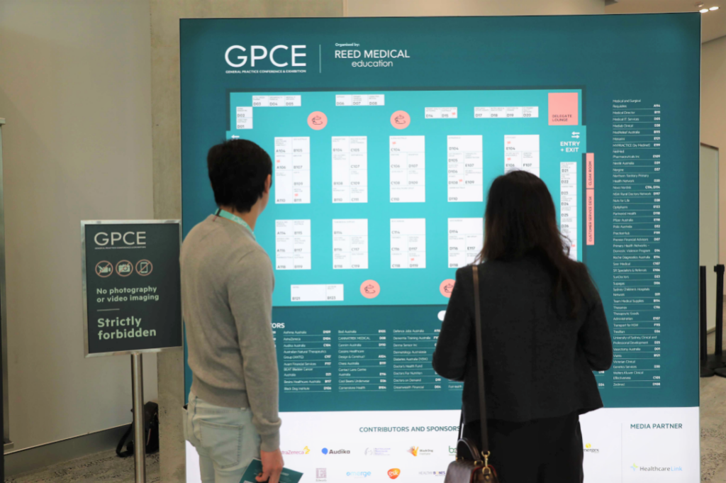
May 15 – 17 | General Practice Conference & Exhibition (GPCE) | Sydney, AUS
GPCE Sydney is one of Australia’s premier educational events for ageneral practitioners and primary healthcare professionals, bringing together clinicians, educators, and industry leaders for three days of independent, CPD-accredited learning and innovation in primary care.
The GPCE welcomes all healthcare professionals working in or with an interest in primary care including GPs, Nurses, Practice Managers, Registrars and Allied Health Professionals.
June 4-6 | European Society of Thoracic Imaging (ESTI) | Salzburg, Austria

The 32nd Annual Scientific Meeting of the European Society of Thoracic Imaging (ESTI) took place June 4–6, 2026 at the Salzburg Congress in Salzburg, Austria.
ESTI’s flagship event brought together radiologists, clinicians, and imaging specialists from across Europe and around the world for a highly engaging in-person scientific program focused on the latest advances in thoracic imaging.
July 17-19 | Florida Radiological Society | Orlando, FL
4DMedical will be attending the 2026 Annual Meeting of the Florida Radiological Society (FRS) and Florida Radiology Business Management Association (FRBMA), taking place July 17–19, 2026, at The Ritz-Carlton Orlando, Grande Lakes in Orlando, Florida. This premier regional meeting brings together radiologists, radiation oncologists, practice leaders, and industry partners to explore the future of imaging and healthcare delivery.
July 31 – August 2 | General Practice Conference & Exhibition (GPCE) | Melbourne, AUS
GPCE Melbourne is one of Australia’s leading events for general practitioners and primary healthcare professionals, offering a dynamic mix of independent CPD-accredited education and an expansive exhibition floor featuring the latest medical innovations and clinical tools. Hosted at the Melbourne Convention and Exhibition Centre (MCEC), this three-day event supports clinicians in expanding their knowledge, sharpening practical skills, and earning valuable continuing professional development hours.
September 12 -13 | General Practice Conference & Exhibition (GPCE) | Brisbane

The GPCE Brisbane 2026 Conference & Exhibition returns as one of Queensland’s key events for general practitioners and primary healthcare professionals, uniting clinicians, practice teams, and industry leaders for two days of accredited education and innovation in primary care.
New Study Underscores XV Technology’s Power to Detect Hidden Lung Disease
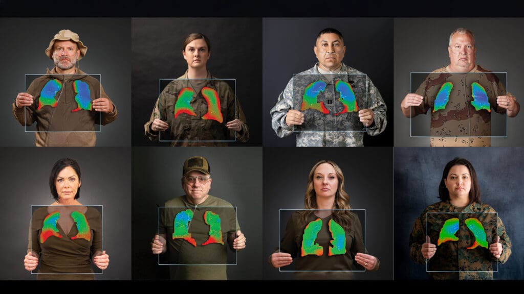
A major new multi-center study published in Respiratory Research (July 2025) demonstrates that 4DMedical’s X-ray Velocimetry Lung Ventilation Analysis Software (XV LVAS®) can reveal early and subtle forms of small airways disease that are often missed by standard tests like spirometry and CT scans. Researchers from Vanderbilt University, Johns Hopkins, University of Miami, and Alfred Hospital in Melbourne showed that XV technology identifies disease-specific and severity-specific biomarker patterns in chronic obstructive pulmonary disease (COPD) and deployment-related constrictive bronchiolitis (DR-CB)—even when conventional tests appear normal.
Innovative XV Technology Shows Promise for Detailed Lung Function Assessment in Bronchiectasis Patients

A new study led by UNSW Sydney researchers demonstrates the feasibility and promise of the innovative XV Scanner™ with XV LVAS®, non-invasive imaging tools developed by 4DMedical, for assessing regional lung function in adults with bronchiectasis caused by Primary Ciliary Dyskinesia (PCD) and other conditions.
Understanding Lung Perfusion: What is it? Why does it matter? And how is it measured?

Lung health is essential for overall wellbeing. A key aspect of healthy lungs is their ability to efficiently transfer oxygen into the blood. This process relies on both the flow of air (ventilation) and the flow of blood (perfusion) through the lungs. While ventilation is about how well air moves in and out, perfusion refers to the movement of blood through the lungs’ fine network of blood vessels. Both are crucial, but today we’ll take a closer look at lung perfusion: what it is, when and why it’s measured, and how advanced imaging technologies are enhancing our understanding of it.
Addressing the Challenge of UIP—and IPF Diagnosis: A Conversation with Dr. Chung
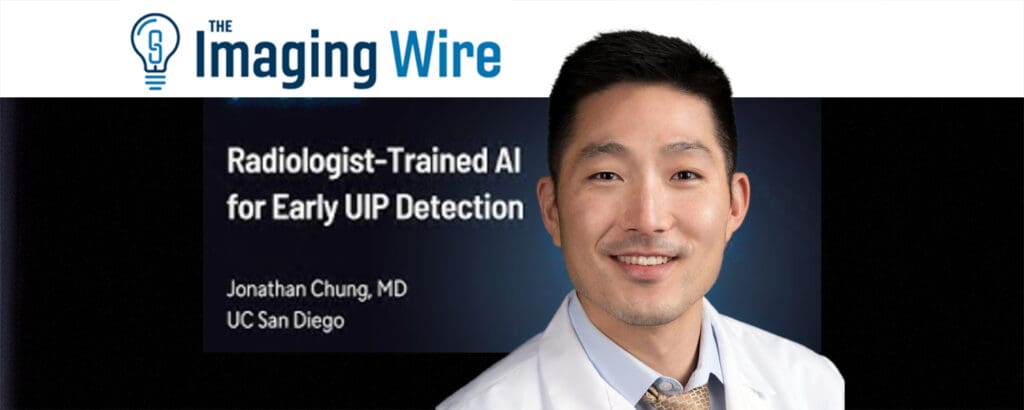
Early identification of Usual Interstitial Pneumonia (UIP) and diagnosis of Idiopathic Pulmonary Fibrosis (IPF) remains a significant challenge in pulmonary medicine. Despite advances in imaging and clinical awareness, the process still requires specialized training and experience that isn’t always readily available—especially in non-urban or resource-limited healthcare settings. This gap can delay diagnosis which critically impacts patient outcomes.
In a recent interview hosted by Brian Casey, editor at The Imaging Wire, Dr. Chung, a leading thoracic radiologist, offers valuable perspective on the complexities associated with UIP identification and the essential role of expert interpretation to achieve accurate diagnoses.
Olympus Launches Emphysema Screening Program Powered by 4DMedical, Expanding Early Diagnosis and Treatment Opportunities

A new peer-reviewed study published in Respiratory Research confirms that 4DMedical’s CT Lung Ventilation Analysis Software (CT LVAS) provides results comparable to those of the gold standard PET method, as well as two additional research techniques for assessing regional lung function. The findings demonstrate a strong association and agreement between 4DMedical’s CT LVAS and PET-ventilation at the lobar level and comparable correlation at the voxel level. Utilizing routine non-contrast CT scans, CT LVAS generates detailed regional ventilation information. This highlights CT LVAS as a safe and contrast-free modality for functional lung imaging, making it an ideal tool for assessing lung function across a range of respiratory conditions.