
CT:VQ® is the world’s first non‑contrast post‑processing technology that transforms routine, chest CTs into quantitative, lobar ventilation (V) and perfusion (Q) maps—without injected contrast or radioisotopes. Delivered via software-as-a-service, results are returned directly into the radiology workflow for interpretation alongside the source CT images.
CT:VQ is FDA-cleared.
Eligible for reimbursement in the US under Category III CPT codes.
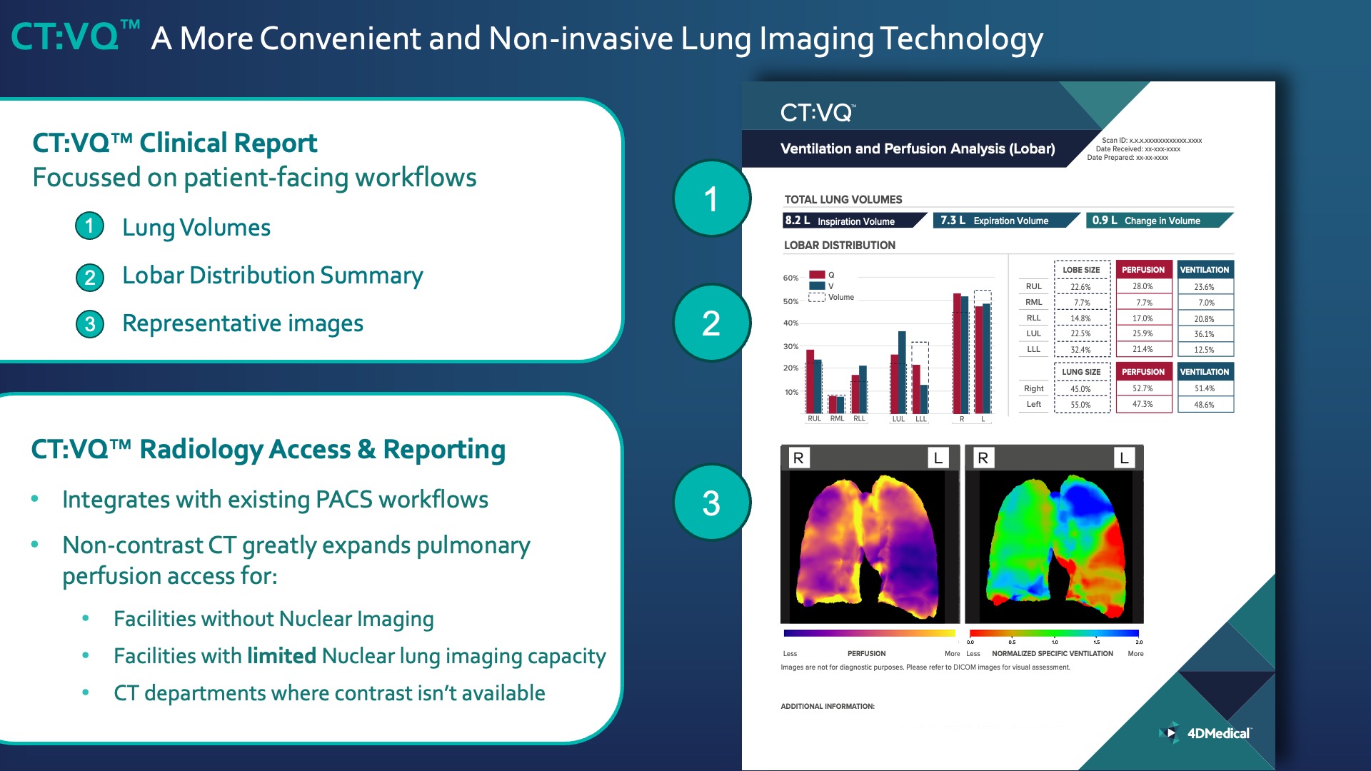
Color V and Q overlays registered to CT anatomy
Quantitative Metrics
Total Lung Volumes (inspiratory, expiratory, and delta)
Lobar Distribution: comparisons between lobe size, ventilation, perfusion, and total right–left lungs
CT:VQ® brings non-contrast functional lung imaging to routine CT and integrates with existing PACS, expanding access even where nuclear imaging or contrast use isn’t available. Its patient-facing report delivers clear, rapid ventilation and perfusion insights—lung volumes, lobar distribution, and representative images.
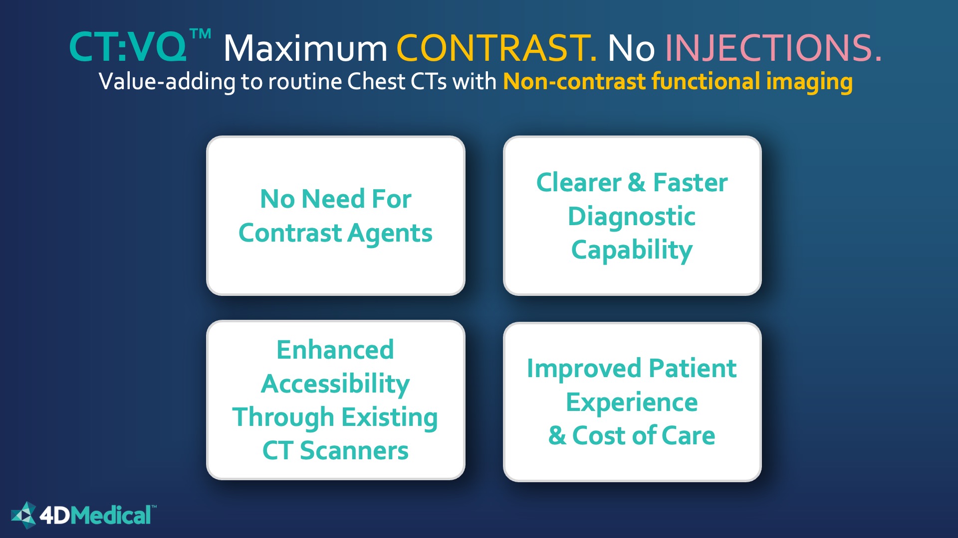
Add functional insights to routine chest CT using existing scanners and PACS—eliminating contrast agents and supporting broader, patient‑friendly access.
CT:VQ® was evaluated using three complementary approaches: Data on file, 4DMedical
1. Quantitative correlation with SPECT V/Q (standalone device performance)
Additional Analysis: CT:VQ® perfusion heterogeneity metrics showed a stronger association with DLCO than comparable SPECT measures in the submission dataset (r²: 0.557 vs 0.380).
2. Reader performance study
3. Case‑based review
Get the latest news about respiratory imaging and ventilation analysis
Please select your region to see the most relevant content and products available in your area.
You are navigating to content specific to another region. Do you wish to proceed?
Availability varies by region. Refer to product labelling for intended use.
We use cookies to improve your experience on our site. By using our site, you consent to cookies.
Manage your cookie preferences below:
Essential cookies enable basic functions and are necessary for the proper function of the website.